
When your vision suddenly changes - like a shadow creeping across your eye or flashes of light that won’t go away - it’s easy to brush it off as eye strain, fatigue, or just getting older. But if you’re experiencing these signs, especially suddenly, you might be facing a retinal detachment. This isn’t a slow-moving issue. It’s an emergency. And every hour counts.
Think of your retina like the film in an old camera. It’s a thin layer of tissue at the back of your eye that captures light and sends signals to your brain. When it pulls away from its normal position, it loses its blood supply. Without that supply, the cells that let you see start to die. And once they’re gone, they don’t come back. That’s why timing is everything.
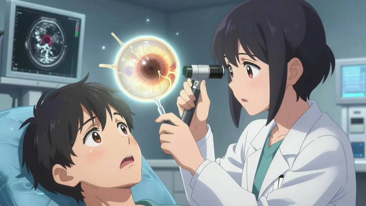
What Does Retinal Detachment Actually Feel Like?
Most people don’t notice anything until it’s too late. But there are six clear warning signs that aren’t normal - and they show up fast.
- Sudden increase in floaters: Not just one or two. You might see dozens of dark spots, squiggles, or cobweb-like shapes that appear out of nowhere. The National Eye Institute says patients often describe it as "a lot of new floaters" appearing within hours.
- Flashes of light: Like someone is snapping a camera in your peripheral vision. These aren’t the occasional sparkles you get when you rub your eyes. These are persistent, especially in dim lighting or when moving your head.
- A dark curtain or shadow: This is the most urgent sign. It feels like a blind spot spreading across your vision - like a curtain being pulled over part of your eye. It usually starts in the peripheral vision and moves inward.
- Sudden blurry or distorted vision: Words on a page look wavy. Straight lines bend. This happens in 68% of cases, according to Cleveland Clinic’s 2021 patient registry.
- Loss of peripheral vision: You might feel like you’re seeing through tunnel vision. This occurs in 73% of cases, as reported by the Retina Research Foundation.
- Changes in color perception: Colors look washed out or duller, especially if the macula - the center of your retina responsible for sharp vision - is involved.
If you have even one of these, especially a curtain or flashes, don’t wait. Don’t call your optometrist tomorrow. Don’t try to sleep on it. Go to the nearest emergency eye clinic or hospital eye department today.
How Do Doctors Diagnose It?
There’s no home test. No app. No over-the-counter remedy. Diagnosis requires specialized tools and training.
The gold standard is a dilated fundus examination. Your doctor will put drops in your eye to widen the pupil, then use an indirect ophthalmoscope - a bright light with special lenses - to look deep into your retina. They’re looking for tears, holes, or areas where the retina has lifted away.
If your eye is cloudy from cataracts or bleeding, they’ll use B-scan ultrasonography. It’s a painless scan that uses sound waves to create a picture of the back of your eye. It’s like an ultrasound for your retina.
And then there’s optical coherence tomography (OCT). This machine takes a detailed cross-section of your retina - like a high-res MRI of the eye. It can show even tiny detachments before they’re visible to the naked eye.
These aren’t tests you get at a regular eye exam. They require equipment like a slit lamp, a 20D or higher indirect lens, and an ultrasound system. That’s why general ophthalmologists miss about 22% of early cases, while retinal specialists get it right 95% of the time.
What Happens If You Wait?
Time is vision.
A 2022 study in the Journal of VitreoRetinal Diseases found that if surgery happens within 24 hours of symptoms, there’s a 90% chance the retina can be successfully reattached. But if you wait 72 hours, your chance of regaining 20/40 vision - the standard for driving - drops from 75% to just 35%.
Dr. Carl Regillo of Wills Eye Hospital says every hour after symptom onset reduces visual recovery by about 5%. That’s not a guess. That’s based on tracking thousands of cases.
One patient on Reddit, "VisionWarrior22," ignored floaters for three days. By the time they went in, the detachment had reached the macula. Their final vision was 20/100. They could’ve had 20/25 if they’d acted sooner.
And here’s another scary stat: 63% of patients in a survey by the American Society of Cataract and Refractive Surgery were initially told by their primary care provider that their symptoms were "eye strain." That delay cost them precious hours.
The Three Main Types of Surgery
There’s no one-size-fits-all fix. The right surgery depends on where the tear is, how big it is, how long it’s been detached, and whether the macula is involved.
1. Pneumatic Retinopexy
This is the least invasive option. It’s done in the doctor’s office. A gas bubble is injected into the eye. You’re then positioned so the bubble floats up and presses against the tear, sealing it. Laser or freezing treatment is used to glue the retina back in place.
Success rate: 70-80% for tears on the upper part of the retina. But it doesn’t work for tears on the bottom. And you have to stay face-down or on your side for 50% of every 24 hours - for up to 10 days. That’s hard. People report headaches, neck pain, and difficulty sleeping.
It’s only for healthy, younger eyes with a single, small tear. About 30% of patients need a second procedure.
2. Scleral Buckling
This is a more traditional method. A soft silicone band is sewn around the outside of your eye. It gently pushes the wall of the eye inward, so it meets the detached retina and holds it in place while it heals.
Success rate: 85-90% for simple cases. It’s often used in younger patients with lattice degeneration - a thinning of the retina that runs in families.
Downside? It can permanently change your vision. On average, it causes 1.5 to 2.0 diopters of nearsightedness. You might need stronger glasses. And 5-8% of patients develop double vision.
3. Vitrectomy
This is the most common surgery today - used in 65% of cases, according to the American Society of Retina Specialists. It’s done in the operating room.
The surgeon removes the vitreous gel - the clear jelly that fills the eye - and replaces it with a saline solution, gas, or silicone oil. This takes pressure off the retina and lets it settle back into place. Laser or freezing is used to seal the tear.
Success rate: 90-95%, especially for complex cases like giant tears or when scar tissue is pulling on the retina.
But here’s the trade-off: 70% of people who have this surgery and still have their natural lens will develop a cataract within two years. That means another surgery down the line.
It’s also the only option if the macula is detached. The 2022 Cochrane Review showed vitrectomy gives better results than scleral buckling when the center of vision is involved.
What to Expect After Surgery
Recovery isn’t quick. And it’s not comfortable.
If you had a gas bubble, you’ll need to position your head exactly as your surgeon says - often face-down. That means eating, reading, and even showering in a specific way. Some people use special face-down chairs. Others tape a tennis ball to their forehead to help them stay in position.
Post-op discomfort is common. A 2022 survey from the Retina Patient Registry found 41% of patients reported significant pain or discomfort from positioning. And 38% needed home health help to manage daily tasks.
Follow-up visits are frequent. You’ll see your surgeon at 1 day, 1 week, and 1 month. They’ll check your eye pressure, look for re-detachment, and monitor for cataracts or infection.
Full healing takes weeks. Vision improvement can take months. Don’t expect perfect sight right away.

Who’s at Risk?
Retinal detachment affects about 1 in 10,000 people each year. But some groups are at much higher risk:
- People with severe nearsightedness (over -5.00 diopters): 167 in 10,000 are affected annually.
- Those who’ve had cataract surgery: 0.5% to 2% risk.
- People with lattice degeneration: 1% lifetime risk.
- Family history of retinal detachment.
- Eye trauma or previous eye surgery.
If you’re in one of these groups, you don’t need to live in fear. But you should have regular dilated eye exams - at least once a year. Catching a tear before it turns into a detachment can prevent surgery entirely.
What’s New in Treatment?
Surgery isn’t standing still.
In January 2023, the FDA approved the EVA Platform by DORC International - a minimally invasive vitrectomy system with 27-gauge tools. Smaller incisions mean less pain, faster healing, and fewer complications.
Surgeons are now using intraoperative OCT during surgery. It gives real-time images of the retina while they’re operating. A 2023 study showed this improved the completeness of scar tissue removal by 15%.
And in the future? Bioengineered retinal patches are in clinical trials. Gene therapies for inherited conditions that weaken the retina are also being tested. These might one day prevent detachment before it starts.
Final Warning: Don’t Ignore the Signs
Retinal detachment doesn’t care if you’re busy. It doesn’t wait for your next appointment. It doesn’t care if you think it’s "just aging."
Floaters? Flashes? A shadow? These aren’t normal. They’re your eye screaming for help.
Act fast. Go to an eye specialist immediately. Don’t wait. Don’t call your GP. Don’t try to wait it out. Your vision - your ability to see your kids, read, drive, live independently - is on the line.
There’s no second chance. But there is a window - and it closes fast.
Can retinal detachment happen in both eyes at the same time?
It’s rare, but possible. Most cases affect just one eye. But if you’ve had a detachment in one eye, your risk for the other eye increases significantly - up to 10-15%. That’s why doctors always check both eyes carefully after one is diagnosed. If you have risk factors like severe myopia or lattice degeneration, regular monitoring is essential.
Is retinal detachment the same as a detached vitreous?
No. A detached vitreous (posterior vitreous detachment, or PVD) is common and usually harmless. It happens when the gel inside your eye shrinks and pulls away from the retina. It can cause floaters and flashes - which are also symptoms of retinal detachment. But PVD doesn’t cause vision loss. The danger is that it can tear the retina as it pulls away. That’s why any new flashes or floaters need to be checked - to rule out a tear or detachment.
Can I prevent retinal detachment?
You can’t prevent all cases, but you can reduce your risk. Wear protective eyewear during sports or jobs with eye hazards. Control diabetes and high blood pressure - both increase eye complications. Get annual dilated eye exams if you’re over 40, nearsighted, or have a family history. If you have lattice degeneration, your doctor might recommend laser treatment to seal weak spots before they tear. But don’t get unnecessary procedures. Not every weak spot needs fixing.
How long does recovery take after retinal detachment surgery?
It varies. Most people can return to light activities in 1-2 weeks. Full recovery takes 2-6 months. Vision improves gradually. If you had a gas bubble, you can’t fly or go to high altitudes until the gas is gone - that can take 6-8 weeks. Cataracts may develop within two years after vitrectomy. You’ll need ongoing eye care, even after healing.
What happens if I don’t get surgery?
Without treatment, the retina will continue to detach, and you’ll lose more vision - permanently. Eventually, the entire retina can detach, leading to total blindness in that eye. The cells that detect light die and don’t regenerate. There’s no medication, eye drop, or natural remedy that can fix it. Surgery is the only option. Delaying it doesn’t just reduce your chances - it makes recovery impossible.
Marcus Jackson
I've seen this before. A friend of mine ignored floaters for 48 hours. By the time he got to the specialist, the macula was already involved. He's lucky he didn't go blind. The stats in this post are real. Every hour matters. Don't be the guy who waits.